flow cytometry results for lymphoma
The goal of the survey which was sponsored by the Clinical Cytometry Society CCS was to document what directors of flow cytometry laboratories currently consider to be. Leukemia and lymphoma analysis by flow cytometry aids in identifying the tumor lineage which in most.

Flow Cytometric Analysis Of Leukemia And Lymphoma The Basics Leukamie
This is especially true if initial testing showed an increased number of.
. This flow cytometry test is used to diagnose leukemia or lymphoma. Immunodeficiency-associated Burkitt lymphoma is usually associated with HIV infection or occurs in the setting of post-transplant patients who are taking immunosuppressive drugs. Not always strictly speaking not very often.
Activation of GSK-3β in CHL results in inhibition of the Wntβ-catenin signaling cascade and its abnormal accumulation in the nuclei of both Reed-Sternberg cells and Hodgkin. Hairy cell leukemia diagnosis. Easy-to-add into multi-color experiments.
In this contest peripheral blood PB and bone marrow BM infiltration have been assessed alternatively by cytology 5-8 flow cytometry FC. The results of a flow cytometry test will show how many irregular cells are present in white blood cells or bone marrow. Depending on specimen type.
Ad NovaFluor dyes designed for spectral flow cytometers. Erythrocytosis with increased hemoglobin is noted on CBC review. Chronic lymphocytic leukemia prognostication.
Stable and minimal spillover. This study examines the reporting of TRBC1 based clonality using flow cytometry and molecular clonality results in the investigation of T-cell lymphocytosis. Flow cytometric leukemia and lymphoma analysis may aid in identifying the tumor lineage for diagnostic and prognostic purposes.
Mycosis fungoides MF and Sézary syndrome SS are the best-studied subtypes of cutaneous T-cell lymphoma a rare non-Hodgkin lymphoma that primarily presents in the skin but can also. The aim of this study was to evaluate the clinicopathological features and flow cytometry FCM of tumor tissues in ocular adnexal diffuse large B-cell lymphoma DLBCL. Flow cytometry is generally used as follow up testing after a complete blood count CBC or white blood cells scan WBC.
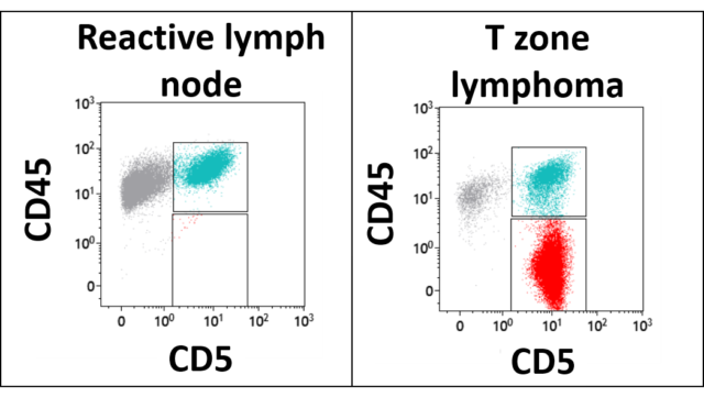
Therefore flow cytometry is an. The results show that four-color flow cytometry improves staging accuracy particularly in patients with a low level peripheral blood or bone marrow involvement. These can be stratified as large and small lymphocytes CD45 positive.
A diagnosis of CLL requires at least 5000 irregular cells. Flow cytometry is usually requested when abnormal cells are seen in the peripheral film. 88184 for the first marker 88185 per marker for each additional marker and 88188 or 88189.
However flow cytometry results usually make certain lymphoma entities extremely likely and others very unlikely. May also include 85060 89050 and 85097. 9-12 or PCR for antigen receptor.
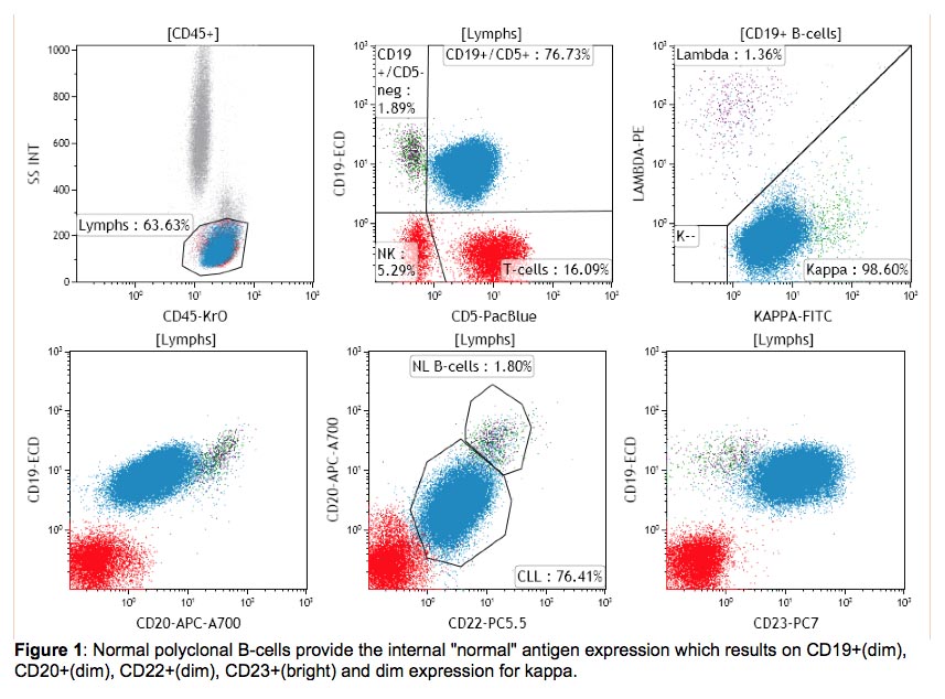
When using fresh tissue for flow cytometric immunophenotyping the predominant populations are lymphoid. After review of the clinical history and morphology a. Ad Learn About A 1L Treatment And See Clinical Data Against A Current Standard Of Care.
With flow cytometry a diagnosis of intraocular lymphoma was confirmed in two of four patients with known lymphoma one of whom had recurrent disease after radiation and not. But there was a stable use of. For me I can instantly tell a lymphoma from a leukaemia just by looking at the film with no flow.
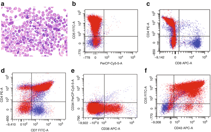
This test is usually done after atypical results are seen on a complete blood count or white blood cell. We compared the antigen expression profile of thymocytes in lymphocyte-rich thymoma with that of precursor T-cell acute lymphoblastic leukemialymphoblastic lymphoma T-cell ALLLBL. Ad Learn About A 1L Treatment And See Clinical Data Against A Current Standard Of Care.
Final interpretation requires correlation with clinical morphologic and other diagnostic information. We have recently demonstrated that classical Hodgkin lymphoma CHL can be immunophenotyped by flow cytometry FC thus obviating the need for immunohistochemistry.

Validation Of A Flow Cytometry Based Method To Quantify Viable Lymphocyte Subtypes In Fresh And Cryopreserved Hematopoietic Cellular Products Cytotherapy

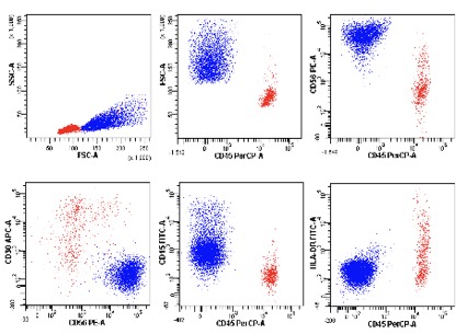
Selected Flow Cytometric Immunophenotyping Plots From Fine Needle Download Scientific Diagram

Flow Cytometry Results Flow Cytometric Graphs Showing Positivity For Download Scientific Diagram
International Clinical Cytometry Society

Flow Cytometric Dot Plots Showing Patterns Of Sig Lcs Negative A B Download Scientific Diagram

Flow Cytometric Immunophenotyping Performed On The Same Plasmablastic Download Scientific Diagram
Principles Of Testing And Publications Clinical Hematopathology Laboratory

Use Of Flow Cytometry In The Phenotypic Diagnosis Of Hodgkin S Lymphoma Grewal 2019 Cytometry Part B Clinical Cytometry Wiley Online Library

Use Of Flow Cytometry In The Phenotypic Diagnosis Of Hodgkin S Lymphoma Grewal 2019 Cytometry Part B Clinical Cytometry Wiley Online Library

International Clinical Cytometry Society
International Clinical Cytometry Society
Flow Cytometry Of Mature And Immature T Cell Lymphoma Springerlink

B Flow Cytometry On Peripheral Blood Revealed An Abnormal Population Download Scientific Diagram
Flow Cytometry Of Mature And Immature T Cell Lymphoma Springerlink

International Clinical Cytometry Society

Flow Cytometric Presentation Of A Large B Cell Lymphoma A Forward Download High Quality Scientific Diagram

Flow Cytometry 3 Acute Lymphoblastic Leukemia Lymphoma What You Need To Know Youtube

Use Of Flow Cytometry In The Phenotypic Diagnosis Of Hodgkin S Lymphoma Grewal 2019 Cytometry Part B Clinical Cytometry Wiley Online Library

Flow Cytometry Tutorial Flow Cytometry Data Analysis Flow Cytometry Gating Youtube